- This topic is empty.
-
AuthorPosts
-
2026-10-10 at 6:03 pm #10272
Assisted reproductive treatment relies on precise, longitudinal hormone data to guide clinical decisions at every stage—from initial evaluation to pregnancy confirmation. Patients entering fertility care often present with irregular or absent periods, infertility, recurrent miscarriage, menopausal symptoms, hirsutism, severe acne, male-pattern baldness, unexplained weight changes, galactorrhea, ovarian cysts, insulin resistance, low libido, erectile dysfunction, gynecomastia, reduced muscle mass, fatigue, depression, osteoporosis unrelated to aging, delayed or precocious puberty, or testicular abnormalities. These overlapping symptoms make it difficult to identify the underlying endocrine cause without a structured testing approach. This is where a comprehensive Sex Hormones panel becomes essential to monitoring during assisted reproductive treatment.
Why Hormone Monitoring Matters During Assisted Reproductive Treatment
A key differentiator in interpreting these symptoms is multi-tier endocrine pathway assessment, which supports separation of central and peripheral dysfunction by evaluating brain centers, pituitary regulators, adrenal glands, gonads, and placental signals. Rather than relying on a single marker, clinicians benefit from a 9-analyte sex hormone profile that supports clinical evaluation of reproductive development, fertility, libido, bone health, metabolic balance, menstrual regulation, and spermatogenesis. Poclight‘s Sex Hormones panel reflects this layered approach, combining nine distinct markers that together map the hypothalamic-pituitary-gonadal-adrenal axis.
Core Markers in the 9-Analyte Sex Hormone Panel
Ovarian Reserve and Gonadotropin Signals: AMH, FSH, LH
AMH (Anti-Müllerian Hormone) functions as an ovarian reserve signal, supporting assessment of ovarian reserve and polycystic ovary syndrome (PCOS) with an AMH >4.7 ng/mL diagnostic threshold. AMH is elevated in PCOS, while it decreases in diminished ovarian reserve and menopause. In males, AMH elevation has no established significance, though it is used to evaluate infant intersex disorders when decreased. For Premature Ovarian Failure, FSH >25 IU/mL combined with AMH <0.5 is diagnostic—illustrating how these markers work together rather than in isolation.
FSH (Follicle-Stimulating Hormone) serves as a gonadotropin reserve signal, supporting evaluation of ovarian insufficiency, menopause, and testicular failure through FSH levels and FSH-based diagnostic ratios. FSH rises in ovarian insufficiency (menopause/POI) in females and primary testicular failure in males, while it falls in hypothalamic/pituitary disorders and hyperprolactinemia.
LH (Luteinizing Hormone) is a gonadotropin status signal that supports distinction between primary gonadal failure and central hypothalamic/pituitary causes when interpreted alongside FSH and sex steroids. LH is elevated in PCOS, ovarian failure, and menopause in females, and in primary testicular failure and Klinefelter syndrome in males; it decreases in hypothalamic/pituitary dysfunction such as Kallmann syndrome. A critical PCOS ratio is LH:FSH ≥2, while Klinefelter syndrome presents with FSH/LH elevation plus decreased testosterone, confirmed by a 47,XXY karyotype.
Estrogen and Luteal Function Signals: E2 and Progesterone
E2 (Estradiol) acts as an estrogen status signal, supporting evaluation of ovarian function, menopausal status, and estrogen-related conditions in both sexes. In females, E2 elevation is associated with ovarian tumors and PCOS, while decreases relate to menopause and ovarian failure. In males, elevated E2 is linked to gynecomastia and testicular tumors, while decreased E2 is linked to hypogonadism. A critical ratio for Premature Ovarian Failure is FSH:E2 >20, making E2 a central value during ovarian function monitoring.
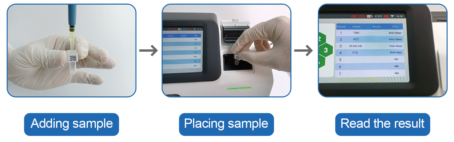
Progesterone provides a luteal function signal, supporting assessment of ovulation and luteal phase adequacy. Decreased progesterone confirms anovulation in PCOS and is the defining feature of luteal phase defect. Elevated progesterone is associated with luteal cysts and molar pregnancy in females, and with adrenal enzyme deficiencies in males. For Congenital Adrenal Hyperplasia, elevated 17-OH Progesterone above 10 ng/mL is diagnostic.
Pregnancy Confirmation and Pituitary Signals: β-hCG and Prolactin
β-hCG (Beta Human Chorionic Gonadotropin) is a pregnancy and trophoblastic disease marker, detecting pregnancy-related conditions and certain germ cell tumors. The clinical interpretation note is unambiguous: always exclude pregnancy before interpreting other hormones in females, and β-hCG >100,000 mIU/mL is diagnostic for Gestational Trophoblastic Disease.
Prolactin (PRL) functions as a pituitary lactotroph signal, supporting detection of prolactin excess—with Prolactin >250 ng/mL suggesting macroprolactinoma. Elevated prolactin is linked to prolactinoma, hypothyroidism, and antipsychotic use, and it correspondingly suppresses LH and FSH, which is why hyperprolactinemia must be ruled out when gonadotropins are unexpectedly low.
Androgen and Adrenal Balance Signals: Testosterone and Cortisol
Testosterone is an androgen status signal, supporting assessment of androgen excess or deficiency in female and male reproductive and metabolic conditions. In females, elevation is associated with PCOS, adrenal tumors, and Congenital Adrenal Hyperplasia, while decrease is tied to hypothalamic amenorrhea. In males, elevation relates to testicular tumors and steroid abuse, while decrease relates to hypogonadism and aging. Klinefelter syndrome is confirmed through FSH/LH elevation combined with decreased testosterone and a 47,XXY karyotype.
Cortisol is an adrenal glucocorticoid marker for stress response and adrenal function, supporting identification of cortisol excess or deficiency through diurnal and threshold-based measures. Elevated cortisol relates to Cushing’s syndrome, chronic stress, and glucocorticoid therapy, while decreased cortisol relates to Addison’s disease and pituitary failure (secondary adrenal insufficiency). Midnight salivary cortisol >145 ng/dL is diagnostic for Cushing’s Syndrome, and AM cortisol <3 μg/dL is diagnostic for Adrenal Insufficiency—both relevant when adrenal dysfunction overlaps with reproductive symptoms.
How the Sex Hormones Panel Supports Treatment Monitoring in Practice
Because these nine analytes map onto brain centers, pituitary regulators, adrenal glands, gonads, and placental signals, clinicians can trace a patient’s symptoms back to a specific point of dysfunction rather than treating markers in isolation. The panel’s delivery/deployment model is standard clinical laboratory testing, with interpretation following local laboratory standards or guidelines, ensuring results integrate smoothly into existing fertility clinic workflows. Its industry adaptation spans reproductive endocrinology, obstetrics/gynecology, urology, endocrinology, pediatrics, and oncology, reflecting the breadth of conditions these nine markers can inform.
This clinical framework is grounded in established reference literature, including Bhasin et al. (2018) on testosterone therapy in men with hypogonadism, the Endocrine Society’s (2011) guidance on diagnosis and treatment of hyperprolactinemia, Husebye, Pearce, Krone, and Kämpe’s (2021) review of adrenal insufficiency in The Lancet, the Rotterdam ESHRE/ASRM-Sponsored PCOS Consensus Workshop Group’s (2004) revised 2003 consensus on diagnostic criteria, and Øksnes and Husebye’s (2023) approach to diagnosing primary adrenal insufficiency in adults.
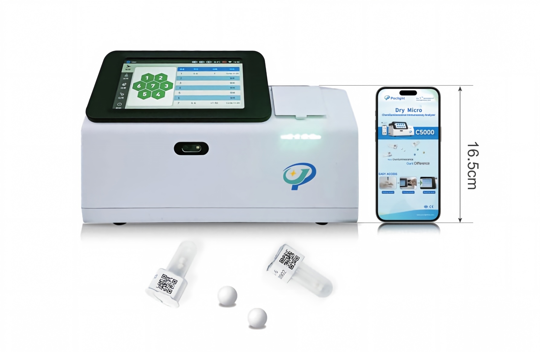
Conclusion
Monitoring during assisted reproductive treatment depends on interpreting hormone values in relation to one another, not as isolated numbers. By combining AMH, FSH, LH, E2, Progesterone, β-hCG, Prolactin, Testosterone, and Cortisol into a single 9-analyte Sex Hormones panel, Poclight supports clinicians in separating central from peripheral causes of reproductive dysfunction, confirming pregnancy status, assessing ovarian reserve, and evaluating adrenal and androgen balance—all within one coordinated clinical laboratory workflow.
http://www.poclight.com
Nanjing Poclight Biotechnology Co., Ltd -
AuthorPosts

- You must be logged in to reply to this topic.